A landmark population study shows near-universal rotator cuff changes with weak symptom correlation, challenging the use of routine imaging in atraumatic pain.
A population-wide imaging study has found that almost every adult over 40 has rotator cuff abnormalities on MRI, regardless of whether they have shoulder pain.
The research, published in JAMA Internal Medicine, has challenged long-held assumptions that structural findings explain symptoms and should guide care.
Melbourne rheumatologist Professor Rachelle Buchbinder was part of the study team, along with researchers from Finland.
“The findings of this study suggest that RC abnormalities are nearly universal after age 40 years and that routine imaging should not guide diagnosis or treatment of atraumatic shoulder pain,” the researchers wrote.
Shoulder pain affects approximately 18% to 31% of the general population globally each month and ranks as the third most common musculoskeletal complaint in primary care, they reported.
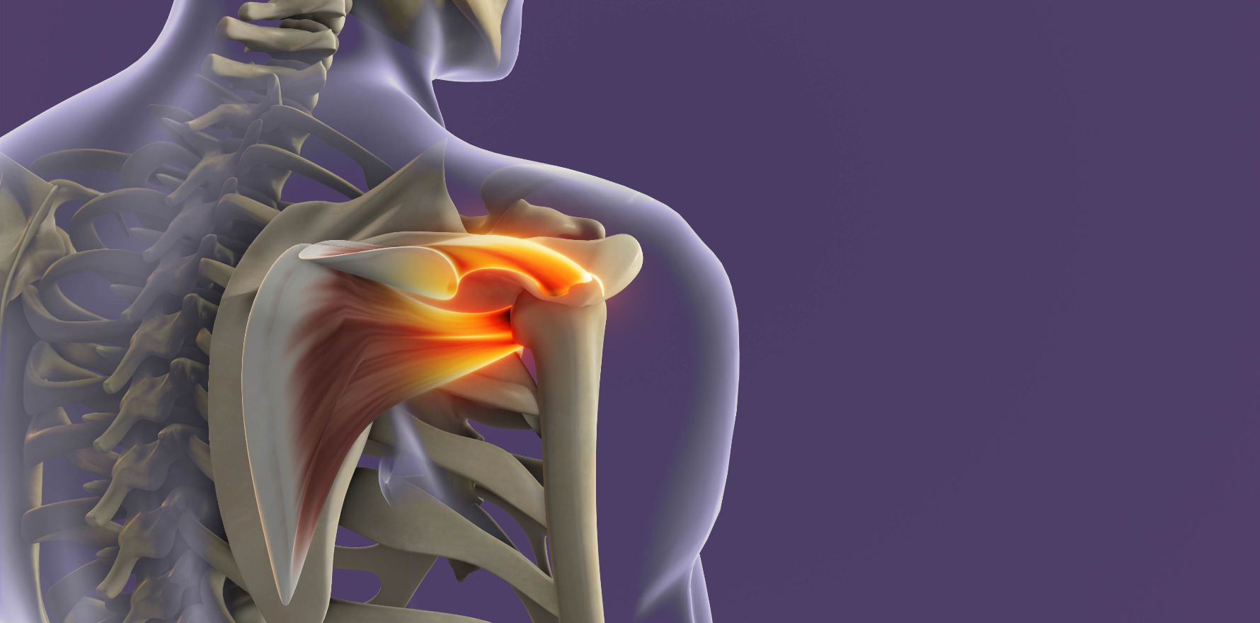
Among individuals presenting with shoulder pain, rotator cuff (RC) abnormalities account for up to 85% of cases.
“Despite a lack of evidence supporting routine use, diagnostic imaging is used in approximately 50% of initial evaluations by general practitioners and considered essential by up to 82% of general practitioners and 56% of specialists,” the researchers wrote.
“Imaging frequently reveals RC abnormalities, such as tendinopathy or partial-thickness tears (PTTs) and full-thickness tears (FTTs).
“Management of these structural findings has become increasingly common and contributes to the growing burden on already strained health care systems.
“In Australia, up to three-quarters of physicians refer patients with shoulder pain to physical therapy, and the use of imaging-guided injections has increased by as much as 46-fold from the year 2000.
“Meanwhile, surgical RC repairs have increased by approximately twofold to sevenfold in other high-income countries.”
In the cross-sectional analysis of 602 Finnish adults aged 41 to 76 years, researchers reported that 99% had at least one rotator cuff abnormality on high-resolution 3-Tesla MRI, with similar rates in asymptomatic and symptomatic shoulders.
The researchers said their findings suggested clinicians should rethink the diagnostic weight given to imaging in atraumatic shoulder pain and reconsider the language used to describe common age-related changes.
“The high prevalence of abnormal MRI findings and their poor concordance with symptoms challenges the routine attribution of shoulder symptoms directly to imaging abnormalities,” they wrote.
“While we cannot dismiss the possibility that some RC tears may contribute to shoulder symptoms, our findings indicate that we are currently unable to distinguish clinically meaningful MRI abnormalities from incidental findings.
“This limitation persists even when using state-of-the-art 3T MRI and conducting detailed clinical assessments by experienced shoulder specialists, highlighting the limited value of imaging and clinical tests for diagnosing RC disorders and guiding treatment decisions.”
Rotator cuff abnormalities were detected in 98.7% of participants, including tendinopathy in 25.3%, partial-thickness tears in 62.4% and full-thickness tears in 11.1%.
The prevalence of abnormalities increased steadily with age but crucially showed minimal association with symptoms. Among more than 1200 shoulders assessed, abnormalities were present in 96% of asymptomatic shoulders and 98% of symptomatic shoulders.
Even full-thickness tears, often considered clinically significant, were frequently silent, with 78% occurring in shoulders without symptoms.
Related


Although unadjusted analyses suggested full-thickness tears were more common in symptomatic shoulders, this difference disappeared after accounting for confounders including other imaging findings and clinical examination results.
The researchers said that such a high baseline prevalence fundamentally altered the diagnostic value of MRI.
When abnormalities are almost ubiquitous, their presence alone carried little specificity or predictive value for pain, making causal attribution unreliable without a clear clinical context.
The new data reinforce concerns about overdiagnosis and overtreatment driven by incidental findings.
The researchers suggested clinicians shift focus from identifying structural “abnormalities” to determining whether imaging findings plausibly explain the patient’s presentation, particularly in the absence of trauma, acute weakness or functional loss.
They also called for more neutral terminology, noting that labels such as “tear” may imply pathology requiring intervention when many findings likely reflect normal ageing. Reframing these changes could reduce patient anxiety and the perceived need for corrective procedures.
Even with state-of-the-art imaging and specialist clinical assessment, the study found clinicians could not reliably distinguish clinically meaningful from incidental findings, underscoring the limits of both imaging and examination in isolation.
“In this cross-sectional study, MRI examination of the shoulder found that RC abnormalities are present in nearly all individuals over 40 years of age, irrespective of symptoms,” the researchers concluded.
“Given that tendinopathy, PTTs, and even FTTs may be incidental findings, clinicians should consider their high population prevalence when interpreting imaging results and deciding on interventions targeting these abnormalities.
“Reframing many of these findings as normal age-related changes rather than disease may help guide more appropriate care and reduce unnecessary interventions. In this context, adopting more precise and less value-laden terminology may help avoid language that implicitly suggests that something is broken and requires fixing.
“Such terminology may reduce patient anxiety, discourage unnecessary surgical interventions and help minimise overdiagnosis and overtreatment.”
