Artificial intelligence isn’t coming to take your job (yet), but it might make a useful assistant someday.
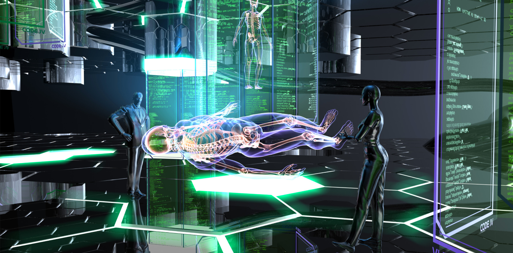
AI will likely be the medical imaging tool of the future, as researchers and early adopters begin to understand its potential uses in identifying synovial swelling, fractures and joint erosion.
The rapid improvements in consumer-facing artificial intelligence models have been regularly making the headlines this past year, but its applications in rheumatology – and healthcare in general – are still unclear.
Speaking at a special EULAR congress session on AI in medicine, Ghent University radiologist Professor Lennart Jans outlined the two broader groups of use cases for AI in imaging: interpretive and non-interpretive.
Both have distinct uses.
Interpretive AI focusses on image interpretation – detecting a fracture, classifying bone age or segmenting vertebra.
The use case here is perhaps what doctors have typically imagined when dreaming up ways to use AI in the consult room.
“Bone age typically takes a long time to determine, but there’s now helpful AI programs for this use,” the Belgian radiologist said.
Essentially, these programs do the same things that a radiologist does, just faster.
Non-interpretive AI, on the other hand, tackles efficiency and productivity issues, like translating radiology reports to layman’s terms, detecting image quality and prioritising certain exams on the worklist.
“A lot of time in our department is wasted, because for every single patient we choose the scanning protocol and so on, but this is no longer the case if you apply AI,” Professor Jans told delegates.
“We can image a lot faster on the MRI if we apply AI tools, and we can scan up to eight times faster for some sequences.”
The obvious upshot is that radiologists, and by extension rheumatologists, would be able to move through patients much faster, thus decreasing hospital waiting lists.
At the Ghent University Hospital where Professor Jans is based, the radiology department uses a non-interpretive AI image translation software to generate synthetic CT scans for some patients.
The result, he said, is an MRI image without radiation.
“Very often with young patients, we really don’t want to put a significant radiation burden on their abdomen, so we … generate a synthetic CT [instead],” Professor Jans said.
Another potential application for AI, according to German rheumatologist Dr Johannes Knitza, is in filling workforce gaps.
He outlined the recent, promising development of a phone application that examines the patient’s hand and detects, via the dorsal skin tone and shape of the fingers, whether the joints are swollen.
“We just do not have enough healthcare professionals, and physical examination is essential,” he told EULAR delegates.
“[If we can now do what] is basically a robot-based ultrasound examination and then an AI model evaluates the images, I think this could become really interesting due to the shortage of rheumatologist healthcare professionals.”
EULAR 2023 was held at the MiCo Convention Centre in Milan, Italy, from 31 May to 3 June.