THE PRESENTATION A 73-year-old lady presented with a five-month history of progressive proximal weakness at the shoulder and pelvic girdles resulting in frequent falls. Other symptoms included swelling around both eyes as well as erythematous macules, on the extensor surfaces of the small joints in her hands and erythema over the V of the neck […]
THE PRESENTATION
A 73-year-old lady presented with a five-month history of progressive proximal weakness at the shoulder and pelvic girdles resulting in frequent falls.
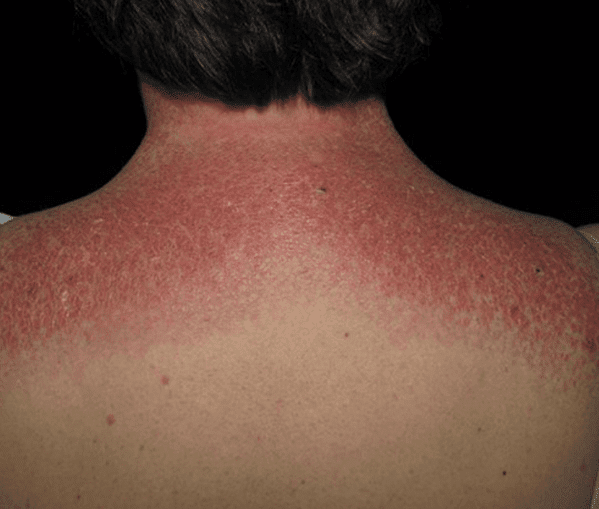
Other symptoms included swelling around both eyes as well as erythematous macules, on the extensor surfaces of the small joints in her hands and erythema over the V of the neck and upper chest.
Further to this she had also experienced dysphagia. There was no history of fever, or dyspnoea, or any new medications.
On general physical examination, her vitals were normal.
She had periungal abnormalities with Gottron’s papules. ([Figures 1 and 2) A violaceous eruption on the upper eyelids and the shawl sign. (Figure 3)
The ear, nose and throat examination were normal.
Neurological examination revealed proximal weakness with grade 3 power at the shoulder and pelvic girdle. Tone and deep tendon reflexes were normal.
Sensory exam was unremarkable. The respiratory, cardiovascular and abdominal systems were all normal.
Her skin biopsy returned showing a non-specific lichenoid pattern, and blood tests found her creatine kinase was elevated to 8317 IU/L. ANA was elevated, 1: 1280 but the ENA was negative.
Myositis specific autoantibodies were tested and Mi-2 was positive.
The patient underwent a contrast CT scan of the neck, chest, abdomen and pelvis, which was unremarkable. To complete her malignancy screen, she went on to have a mammogram, gastroscopy and colonoscopy which were all negative.
After a planning soft tissue MRI; she went on to have a muscle biopsy which was diagnostic for dermatomyositis.

QUICK FACT CHECK
Dermatomyositis is an idiopathic inflammatory myopathy. There is a female to male predominance of about two to one.1 The peak incidence in adults occurs between 40 to 50 years, however individuals of any age can be affected. Estimates of prevalence range from five to 22 per 100,000.2
The incidence of malignancy is high, particularly in the older age groups. Diagnostic criteria include typical cutaneous features, progressive proximal symmetrical muscle weakness, elevated muscle enzymes and pathognomonic findings from muscle biopsy.
BACK TO THE CASE
Initial treatment comprised of pulse intravenous methylprednisolone 1g daily for three days along with induction intravenous immune globulin.
She was then started on oral prednisolone 50mg daily on a tapering course and methotrexate was added as a steroid sparing agent.
She recovered well, her rash faded, her creatine kinase normalised and she regained muscle strength gradually. Her prednisolone was tapered to a maintenance of 5mg daily and she continued on monthly intravenous immune globulin.
RELAPSE
Eleven months after the onset of her illness, her disease relapsed.
She noted a return of her rash and proximal weakness. Her creatine kinase rose to 6500 IU/L. Therapy was escalated and she had a second cycle of pulse intravenous methylprednisolone and re-induction intravenous immune globulin.
Intensification of treatment led to other complications including worsening control of her type II diabetes and fatty liver disease.
Her dermatomyositis eventually went into disease remission, however she developed multiple erythematous nodules over her abdomen. (See Figure 4, page 9)
A deep biopsy was taken of the lesion and sent for histopathology and culture.
Histopathological examination showed an inflammatory infiltrate, and poorly formed granulomas.
Mycobacterium chelonae was grown from the specimen cultures and was identified by high performance liquid chromatography and line probe assay.
Susceptibility testing showed susceptibility to cefotixin; intermediate susceptibility to linezolid, tobramycin and amikacin.
Dual therapy with cefotixin and linezolid was commenced. The patient developed sensitivity to cefotixin and this was ceased.
Tobramycin was started with a deterioration of renal function.
The decision was made to manage the lesions locally with surgical excision with control of the infection.

DISCUSSION
Dermatomyositis is characterised by an erythematous rash and symmetric proximal muscle weakness. Muscle biopsy reveals a mononuclear, inflammatory cell exudate arranged in a perivascular and perifascicular distribution with degenerating and regenerating muscle fibres and perifascicular atrophy.
Emerging data suggests that the autoantibody status of patients with idiopathic inflammatory myopathy defines phenotypes and predicts outcomes; this is found in the serum of 50 to 60% of patients.3
In this case, MI-2 was identified; it is directed against a helicase involved in transcriptional activation.4
Among patients with dermatomyositis, anti-Mi-2 antibodies are present in about 7% of Caucasians. It is associated with the relatively acute onset of disease, traditionally associated with a classic shawl or V sign, and may respond well to therapy.5
The approach to treatment of dermatomyositis has not been standardised because of the rarity of the disease and lack of controlled treatment trials.
Despite the absence of placebo-controlled trials demonstrating their effectiveness, glucocorticoids remain key in initial therapy. Concurrent with glucocorticoids, an immunosuppressive drug is added to function as a steroid sparing drug; the usual choices are methotrexate, azathioprine and mycophenolate.
No trials have shown the superiority of one of these agents over others.3
In cases where the disease is severe or the patient presents with dysphagia and is at risk of aspiration; intravenous immune globulin is added early, as it may have a more rapid onset of action. Another advantage is that it is not an immunosuppressant, however prolonged treatment is limited by difficulty of administration and cost.
Therapy for more refractory cases include cyclophosphamide, rituximab and cyclosporine A.
Immunosuppressed patients are vulnerable to opportunistic infections and there should be a high index of suspicion.
In this case, the patient developed multiple Mycobacterium chelonae abdominal abscesses as a complication of her immunosuppression. Mycobacterium chelonae is a rapidly growing, non-tuberculous mycobacterium. A ubiquitous microorganism, it is found in soil, water, sewage and dust particles.
In humans, the organism is an uncommon cause of localised cutaneous lesions (e.g. associated with surgical wounds or acupuncture) and also in disseminated disease.6 Often the source is contamination from colonised water tap. In this case, the needle from the patient’s insulin pen was suspected to be the culprit. However, culture of the needle returned negative.
Treatment for Mycobacterium chelonae infection is challenging, as the organism is resistant or only partially susceptible to many antibiotics. To avoid the emergence of resistance, dual treatment is recommended.7
Therapy needs to be continued for several months which can be difficult.
In the case described here, surgery was chosen ultimately as the management option for local control of the abscesses. (Figure 5)
Declaration of patient consent: Informed consent was obtained from the patient for publication of this manuscript and any accompanying images.
Dr Anne Chung, MBBS, FRACP, is a consultant rheumatologist based in Parramatta, Sydney
References:
1. Dermatopolymyositis and other connective tissue diseases: a review of 105 cases. Tymms KE, Webb J, J Rheumatol. 1985;12(6):1140.
2. Estimating the prevalence of polymyositis and dermatomyositis from administrative data: age, sex and regional differences. Bernatsky S, Joseph L, Pineau CA, Bélisle P, Boivin JF, Banerjee D, Clarke AE. Ann Rheum Dis. 2009;68(7):1192.
3. Idiopathic Inflammatory Myopathies: Current Trends in Pathogenesis, Clinical Features, and Up-to-Date Treatment Recommendations. Floranne C. Ernste, MD, and Ann M. Reed. Mayo Clin Proc. January 2013;88(1):83-105
4. The major dermatomyositis-specific Mi-2 autoantigen is a presumed helicase involved in transcriptional activation. Seelig HP, Moosbrugger I, Ehrfeld H, Fink T, Renz M, Genth E Arthritis Rheum. 1995;38(10):1389.
5. A new approach to the classification of idiopathic inflammatory myopathy: myositis-specific autoantibodies define useful homogeneous patient groups. Love LA, Leff RL, Fraser DD, Targoff IN, Dalakas M, Plotz PH, Miller FW Medicine (Baltimore). 1991;70(6):360.
6. Nontuberculous mycobacterial infections: a clinical review. Wagner D, Young SL. Infection 2014; 32: 257 – 270.
7. Cutaneous Mycobacterium cheonae-abscesses. Grandinetti L, Tomecki K et al. J Cutan Pathol 2007; 34: 88


